Cyanotic heart diseases in dogs: when erythrocytosis becomes problematic
£25.00 + VAT
SKU: CAR-ERYTH

Add up to 3 PDFs to your basket together with an Online Course, and get the PDFs for FREE!!!
Overview
Cyanotic heart diseases in dogs: when erythrocytosis becomes problematic
The paper reviews the diagnostic approach to erythrocytosis in dogs, with a focus on cyanotic congenital heart disease and management of associated complications.
Congenital heart diseases such as pulmonic stenosis, subaortic stenosis, and patent ductus arteriosus are usually identified through loud murmurs and echocardiography. More complex congenital heart diseases are less common and may not produce obvious murmurs, delaying diagnosis until complications such as erythrocytosis develop.
Erythrocytosis is defined as an increased packed cell volume or haematocrit above 55% that is not caused by dehydration. Clinical signs usually occur when haematocrit exceeds 70–75%, producing hyperviscosity syndrome. Consequences include sluggish capillary blood flow, microthrombi formation, muscle pain, exercise intolerance, seizures, and neurological signs.
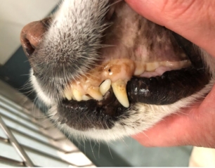
Cyanosis develops when circulating deoxyhaemoglobin reaches sufficient concentration to produce blue-grey mucous membranes. Dogs with erythrocytosis may appear cyanotic at lower levels of respiratory compromise because of increased deoxyhaemoglobin concentration.
Erythrocytosis may be primary or secondary. Primary erythrocytosis is usually caused by polycythaemia vera. Secondary erythrocytosis results from increased erythropoietin production in response to hypoxaemia. Investigation of erythrocytosis should include assessment for cardiovascular disease, respiratory disease, renal disease, and neoplasia.
Congenital cardiac diseases associated with erythrocytosis typically involve a right-to-left shunt that allows deoxygenated blood to enter systemic circulation. Conditions discussed include tetralogy of Fallot, atrioventricular septal defects, atrial septal defects with pulmonic stenosis, patent foramen ovale, right-to-left patent ductus arteriosus, and pulmonary hypertension.
Pulmonary hypertension is described as increased resistance to pulmonary blood flow resulting in elevated right ventricular afterload. Causes include congenital heart disease, chronic mitral valve disease, respiratory disease, thromboembolic disease, and parasitic disease. Echocardiography with Doppler assessment is considered the primary diagnostic method.
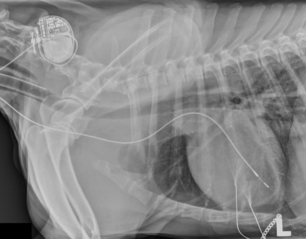
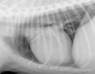
Several clinical case examples are presented. One French Bulldog with tetralogy of Fallot and severe erythrocytosis underwent a modified Blalock-Thomas-Taussig shunt procedure, resulting in reduction of haematocrit from 73% to 46%. A Jack Russell Terrier with atrioventricular septal defect and pulmonary hypertension was managed medically with sildenafil and haematocrit control. An English Bulldog with severe pulmonic stenosis and atrial septal defect underwent balloon valvuloplasty and atrial septal occluder placement, reducing haematocrit from 79% to 41%. A Cavalier King Charles Spaniel with pulmonic stenosis and patent foramen ovale underwent balloon valvuloplasty with subsequent closure of the shunt and normalisation of haematocrit.
The article explains tetralogy of Fallot as a combination of ventricular septal defect, overriding aorta, pulmonic stenosis, and right ventricular hypertrophy. Historical development of the Blalock-Taussig shunt procedure is also discussed.
Management of erythrocytosis depends on the underlying cause and severity of clinical signs. In dogs with right-to-left shunting, erythrocytosis is considered physiologically appropriate because increased red blood cell mass helps maintain tissue oxygen delivery. Treatment aims to balance oxygen-carrying capacity with reduction of hyperviscosity complications.
Target haematocrit is approximately 65%, while levels above 75% generally require intervention. Sildenafil is recommended for cases associated with pulmonary hypertension. Phlebotomy may be used for emergency or maintenance management of severe erythrocytosis, with simultaneous crystalloid replacement. Hirudotherapy using medical leeches is presented as an alternative method for smaller patients. Hydroxyurea may be used when repeated phlebotomy is poorly tolerated or impractical.
The paper concludes that dogs with complex congenital heart disease and erythrocytosis can often achieve prolonged survival and good quality of life with appropriate monitoring and management, even when definitive surgical correction is not possible.
 This is a PDF of an article published in VetCPD Journal. For the most current CPD we highly recommend our range of 70 Online Courses – see below for details. Please note that these articles do NOT have exams associated with them.