Haematology testing is characterised by automated analysis performed with a dedicated analyser and accompanied by blood smear examination. The latter can provide crucial clinical information that might otherwise be missed if this step is skipped. Let’s explore five reasons why blood smear examination is essential:
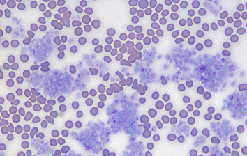
- Platelet Clumping: the most common cause of thrombocytopaenia in dogs and cats is platelet clumping. This spurious change can be flagged by some analysers, but examining the blood smear for the presence of clumps on the feathered edge represents the most accurate method of assessment (see picture above). Without this step, cases of thrombocytopaenia could be significantly overestimated.
- Red Blood Cell Morphology: morphological changes affecting red blood cells are often critical for diagnosing anaemia and determining its cause. For instance, the presence of numerous spherocytes in a dog with regenerative anaemia would strongly suggest immune-mediated haemolytic anaemia (IMHA).
- Haemoparasites: although it is uncommon to identify haemoparasites in blood smears, when they are detected, a diagnosis can be made without the need for further tests such as PCR. These include pathogens such as Babesiaspp., Ehrlichia spp., Anaplasma spp., and Mycoplasma spp. These pathogens cannot be confirmed through instrumental analysis alone.
- Toxic Changes / Band Neutrophils: one of the primary indicators of inflammation is the presence of neutrophilia accompanied by band neutrophils (immature forms with a U-shaped nucleus) and toxic changes (increased cytoplasmic basophilia, Döhle bodies). These features can be easily appreciated via blood smear examination but are not confirmed through automated analysis. In fact, when present, the analyser may provide incorrect leukocyte differential counts, often indicating spurious neutropenia and lymphocytosis.
- Atypical Cells / Leukaemia: leukaemias are neoplasms affecting circulating haematopoietic cells and involving the bone marrow and peripheral blood. They are often, but not always, accompanied by significant leucocytosis. A definitive diagnosis requires evaluation of cell morphology, which is possible with blood smear examination, and further characterisation usually involves immunophenotyping procedures such as flow cytometry. Automated analysers in these cases record leucocytosis but cannot provide a differential count due to these morphological changes.