Cytology is often a first-line diagnostic test that can frequently provide a diagnosis in cases of localized or generalized lymphadenomegaly. However, samples can sometimes be non-diagnostic for various reasons. Let’s look at three common mistakes in lymph node cytology and how to avoid them:
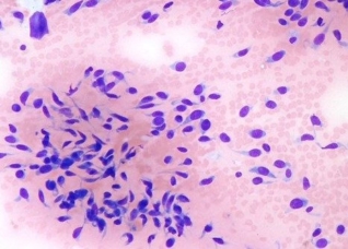
• Sample the lymph node (not the salivary gland): It may sound obvious, but it is not uncommon to accidentally sample a salivary gland when attempting to aspirate the mandibular or submandibular lymph node. Ultrasound-guided sampling can be helpful in these situations. Have a look at the vacuolated salivary gland cells in the image on the left—an example of a misdirected sample. In cases of generalized lymphadenomegaly, don’t limit yourself to the submandibular lymph node just because it’s the easiest to reach. It is important to sample multiple enlarged nodes to obtain a representative picture of the process.
• Don’t make the smear too thick: Lymph nodes exfoliate well, often yielding a high number of cells. Ensure your smear is evenly spread across the slide with cells arranged in a monolayer. Otherwise, you may end up with slides where the cells are poorly stained, overlapping in multiple layers, and difficult or impossible to evaluate—like the example shown in the center image.
• Respect the cells: Lymphoid cells are relatively fragile compared to other cell types. Excessive negative pressure during aspiration or pressing too hard when smearing can damage the cells, resulting in the presence of bare nuclei, as shown in the image on the right. Note how most cells appear as pale purple aggregates of nuclear material with indistinct borders. To avoid this, consider using the needle-only technique (without a syringe), as lymphoid cells exfoliate easily. Be gentle during the smearing process. You may also want to stain and check the slides before submission—if most of the cells are damaged, a non diagnostic will not be likely.