Cytology is often a first-line diagnostic test that can frequently provide a diagnosis in a short period, in a non-invasive manner, and at a limited cost. However, samples can sometimes be non-diagnostic due to low cellularity, poor preservation, or hemodilution. Let’s look at five tips that may help increase the chances of obtaining a diagnostic sample:
- Choose the correct tools: Use needles, typically 22-25 gauge, to collect samples. The larger the needle, the more material you will collect but the higher the chance will be to cause hemodilution. Large needles may be preferred for solid /hard lesions that may poorly exfoliate (e.g. sarcoma like lesions). Small needles may be preferred for organs that exfoliate very generously (e.g. lymph nodes). If you decide to apply suction to increase the cell exfoliation, you may want to select a syringe that is 2 to 5 mL to provide adequate suction without excessive force, which may result in cell damage. Smears should ideally have rounded borders and a frosted end, allowing you to write patient details on it with a pencil.
- Choose the correct sampling procedure: Assess the lesion or organ thoroughly to decide the best approach for sample collection. For hard lesions that may poorly exfoliate, prefer a fine needle aspiration (FNA) with a syringe. For lesions that are soft, vascularized, and/or have a high tendency to exfoliate, such as lymph nodes, the needle-only technique is preferred. If the lesion is very thin and ulcerated, an impression smear may be used instead. In this scenario, please be aware that the sample collected will likely be representative only of the superficial part of the lesion.
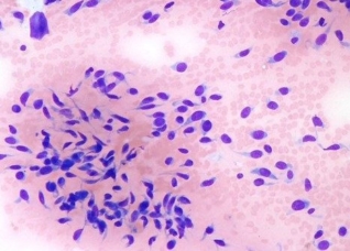
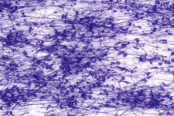
- Prepare a correct smear via squash preparation technique: This is the technique most commonly used in cytology and provides the best results. Attention must be paid to applying the right amount of pressure when a second slide is placed over the slide with the material. Excessive pressure may result in cell rupture, as shown in the picture above, where nuclear streaming prevails. The two slides should then be quickly and smoothly pulled apart to create a thin, even smear. Smears that are too thick, with cells arranged in multiple layers, may not be diagnostic.
- Optimize the staining procedure: Rapid stains such as Diff-Quik are the most commonly used in veterinary practices. Attention must be given to performing the procedure correctly (refer to the stain datasheet) and to regularly changing the stains. Giemsa-based stains may produce better results, especially in terms of nuclear details, which are important for the diagnosis of neoplasia, and for staining mast cell granules.
- Use a good microscope: It is important that the microscope you use for examination is in good condition and has clean lenses. Ensure regular maintenance, including cleaning the lenses and calibrating the light source.